About the Reproductive Sciences and Regenerative Medicine Unit
Core Scientists in the Reproductive Sciences and Regenerative Medicine Unit contribute to the Primate Center mission through NIH-supported programs and peer-reviewed publications; by serving the greater research community in Cores, NIH-supported Centers, and outreach programs; by enhancing the nonhuman primate resource with new assays, biomarkers, model development, and innovative in vivo imaging paradigms; through participation in the management of the reproductive colony and Primate Center administrative positions and committees; and by mentoring trainees and investigators at all career stages in the use of the nonhuman primate as a translational model. Unit Core Scientists serve as collaborative hubs and have outstanding track records in forming multidisciplinary partnerships and teams as evidenced by extramural grants, key publications, and leadership positions in UC Davis and national centers and programs. These include the UC Davis Clinical and Translational Science Center (CTSC), the School of Medicine Stem Cell Program and Gene Therapy Center/Institute for Regenerative Cures, Center for Molecular and Genomic Imaging, and Center for Health and the Environment. Unit Core Scientists bring their extensive expertise and strong track records to partnerships in gamete biology and reproductive toxicology; regenerative medicine and stem cell transplantation; gene therapy and genome editing; lifespan health–from the earliest developmental stages to aging populations; in vivo imaging tools and technologies for translational research; and the conduct of investigational new drug (IND)-enabling studies for human translation. The depth and breadth of expertise, services, and accomplishments contribute substantially to the Primate Center mission, significantly enhance the resource, and ensure that investigators nationwide can conduct state-of-the-art investigations with nonhuman primates at the highest quality level, and with the essential rigor and reproducibility.
Focus on Lifespan Health. Monkeys and humans share many reproductive and developmental features that emphasize their importance as translational models. The unique expertise in the Unit provides a means to address research questions associated with all developmental stages (embryo, fetus, newborn, infant), juveniles; young adults; pre-menopausal/transitional reproductive stages; and advanced geriatrics.
Preclinical and IND-Enabling Studies
Research Accomplishments in Reproductive Sciences and Regenerative Medicine
- The preimplantation period encompasses restructuring and remodeling of the embryonic genome and reprogramming of gene expression. RNA-seq libraries, sequencing, and systems analysis were incorporated to discover the underlying mechanisms that drive major changes in gene regulation during preimplantation development in rhesus monkeys.
- Developing new intravaginal women-controlled, non-hormonal contraceptive devices that also have antimicrobial action. Studies also characterize how these devices alter the normal vaginal microbiome during various phases of the menstrual cycle.
- Demonstrated that ovarian steroids can change both the structure and function of the adrenal glands, which may explain differences in menopausal symptoms and health trajectories in middle-aged women.
- Bisphenol A (BPA) is an endocrine disrupting compound that is an environmental contaminant. BPA-associated alterations were shown in the fetal oviduct by Affiliate and Core Scientists, and such changes could potentially affect oviduct morphology and function later in life with an impact on fertility.
- Studies by Core Scientists across two Research Units (with Cardiorespiratory Diseases) addressed the impact of environmental exposure to wildfire smoke during conception and pregnancy on animals housed outdoors. These studies suggested a possible relationship between air pollution exposure and an increase in pregnancy loss, specifically fine particulates that are relevant to human exposure.
- Studies characterized the transcriptional landscape of the developing rhesus monkey retina allowing a comprehensive understanding of foveal development, with several key miRNAs differentially expressed in the presumptive fovea. Findings identified new ways to direct pluripotent stem cells towards cone photoreceptors.
- Investigations addressed microglial cells as an integral component of cortical proliferative zones during fetal development and the interactive milieu critical to development. These studies used fetal intraventricular gene transfer under ultrasound guidance in early gestation to label the cells.
- Biopharmaceuticals are often PEGylated to enhance their pharmacokinetics. However, recent clinical trials have demonstrated that administration of PEGylated compounds can induce anti-PEG antibodies. Results of studies with rhesus monkeys showed that anti-PEG antibodies can limit the activity of PEGylated drugs and compromise activity of otherwise effective therapeutic agents. These findings have importance for drug/vaccine development and nonviral gene editing tools that are PEGylated.
- Extensive expertise with the fetal rhesus cytomegalovirus (RhCMV) model was leveraged for NIH funding to address Zika virus teratogenesis and trafficking. In one study, while typical fetal growth and development were observed, significant changes were noted in the developing brain at three weeks post-fetal Zika infection including alterations in the distribution, density, number, and appearance of microglial cells, the immune cells of the brain. In addition, the distribution of neural precursor cells was found to be impacted. At three months post-infection, similar changes were observed, which was accompanied by sustained maternal immune system activation. Other studies with Core Scientists at the Wisconsin NPRC have focused on sexual transmission of Zika virus and pregnancy.
- Studies have also elucidated the development of innate-memory T cells, with mixed innate and adaptive features, in the context of RhCMV infection of infant rhesus monkeys. This work is significant because it uncovers an important sequel to CMV infection in childhood, which helps to establish the inflammatory setpoint that partly determines disease susceptibility. The inflammatory setpoint has been an ongoing area of research interest for Unit Scientists.
- In relation to lifespan health, studies examined the impact of CMV on the adult microbiota and immune responses. This work addressed the confounding heterogeneity in immune responses of older individuals to vaccines and infections, which was found to be rooted in the presence or absence of persistent CMV.
- Established novel in vivo imaging methods to monitor gene expression long-term, and showed high levels of reporter gene expression with no adverse effects up through ~16 years of age when organ-targeted fetal gene transfer approaches were used.
- Synthesized radioimmunoconjugates using radioactive copper (64Cu) and zirconium (89Zr) for cell trafficking, and optimized radiolabeling methods to identify engrafted human cells in the rhesus host.
- Showed multilineage engraftment after fetal transplantation of cytokine expanded human cord blood CD34+ cells. These findings are significant because they have shown human cells in the rhesus host that persist over time and without pre-transplant conditioning. Bioluminescence imaging has revolutionized monitoring engraftment and the induction of tolerance to transplanted cells.
- Core Scientists designed and optimized performance of an innovative total-body PET scanner (EXPLORER) that has been commercialized and shown high levels of sensitivity in nonhuman primates and human subjects (see Multimodal Imaging Core).
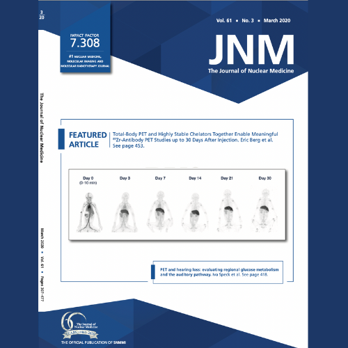
- Demonstrated zirconium (89Zr)-radiolabeled antibody biodistribution out to 30 days post-injection, allowing a better understanding of antibody behavior than previously observed and aided in identifying chemically stable chelator moieties appropriate for use out to these unprecedented imaging timepoints. This publication was selected in 2021 as the Best Overall Paper published in 2020.
Primate Center for Gene Therapy
The Primate Center for Gene Therapy provides collaborative opportunities to address crucial gaps, and accelerate new NIH grant submissions and IND applications. Read more.
Building on extensive expertise in gene therapy and the deep experiences in the Gene Therapy Center, the Nonhuman Primate Testing Center for Evaluation of Somatic Cell Genome Editing Tools was established to develop safe and effective somatic cell gene editing methods to treat patients with common or rare diseases. The Testing Center supports a range of projects targeting a variety of cells and organ systems. The Somatic Cell Genome Editing (SCGE) Consortium includes 72 PIs, 45 projects, and 38 institutions across the U.S. to develop targeted systems for the delivery of new editors and improved human genome editing tools as well as exploring new methods for assessing unintended biological effects. [Link]
Translational Human Pluripotent Stem Cell Shared Research Facility (TSRF)
The UC Davis Stem Cell Program combines unique capabilities for stem cell research with training in diseases that might be prevented, reversed, or ameliorated by stem cell therapy. The TSRF is a state-wide service facility dedicated to the study of human stem and progenitor cells. The facility includes cell culture laboratories, flow cytometry and cell sorting, a molecular core, a histology core, controlled-rate cryopreservation and cell storage for cell lines and cell banks, and an infrastructure of experienced personnel to ensure efficient operation, to provide services, and for training and research guidance (www.tsrf.ucdavis.edu). To learn more about the TSRF, download a pdf here.